How to Identify Constricted Ear Deformity in Newborn Infants
Constricted ear deformity, often referred to as cup ear or lop ear, appears when the upper rim of the newborn’s ear is tightly folded or wrinkled. Parents and healthcare providers can identify this condition through visual inspection for a smaller and flatter ear that may seem tethered to the side of the head. Palpation can also help assess how firm and structured the ear is. It’s important to distinguish constricted ears from helical compression, which has all necessary skin and cartilage present. Early recognition is essential for effective treatment, making resources like Ear Well Centers crucial for providing specialized care that enhances outcomes without surgery.
1. Ear Well Centers of Excellence for Ear Deformities
Ear Well Centers of Excellence play a crucial role in the treatment of constricted ear deformity. These specialized centers offer non-surgical approaches, such as the EarWell Infant Ear Correction System, designed to reshape malformed ears gently. This method is particularly effective in cases where the ear is not fully formed, as it encourages the growth of missing skin and cartilage, leading to a more natural ear shape.
The centers are staffed by experts who have conducted significant research into ear deformities. Specialists like Dr. Steve Byrd and Dr. Eric Payne are known for their innovative techniques that often result in better outcomes than traditional surgical methods. They focus on individualized care, tailoring treatment plans to each infant’s specific needs.
In addition to treatment, these centers provide continuous follow-up care, ensuring that the EarWell device is adjusted as needed for optimal results. Parents receive educational resources to help them understand their child’s condition and the treatment process, fostering a supportive environment for both the child and the family.
2. What is Constricted Ear Deformity?
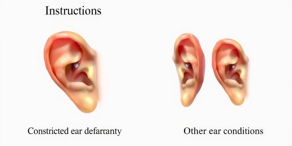 Constricted ear deformity, often referred to as cup ear or lop ear, is a condition where the upper rim of the ear, known as the helical rim, appears tightly folded or wrinkled. This deformity indicates that certain parts of the ear are not fully formed, which may include missing skin or cartilage. The ear may look smaller and flatter than a typical ear and can sometimes appear to be pulled toward the head. Identification of this condition can be critical for early intervention, as it may affect the child’s overall appearance and self-esteem as they grow. Understanding the characteristics and implications of constricted ear deformity is essential for parents and healthcare providers.
3. How to Identify Constricted Ear Deformity
Constricted ear deformity, commonly referred to as cup ear or lop ear, is identified mainly through visual inspection. Parents and caregivers should look for key characteristics: a helical rim that appears folded or wrinkled, a smaller and flatter overall appearance of the ear, and the ear potentially appearing tethered to the side of the head. It’s important to differentiate this condition from helical compression, which retains all skin and cartilage and usually corrects more easily. A gentle palpation of the ear can also help assess its firmness and structure, providing further insights into the deformity. Early identification is essential for effective intervention, as prompt treatment can lead to better aesthetic outcomes and may prevent the need for surgical correction later in life.
| Identification Method | Description |
| — | — |
| Visual Inspection | Look for characteristics such as a folded or wrinkled helical rim, smaller and flatter appearance of the ear, and tethered appearance to the side of the head. |
| Differentiation from Other Conditions | Distinguish between constricted ear deformity and helical compression. |
| Palpation | A gentle examination to assess firmness and structure of the ear. |
4. Differentiating Constricted Ear Deformity from Other Conditions
To accurately identify constricted ear deformity, it’s important to differentiate it from other ear conditions. One common condition is helical compression, where the ear also appears folded but retains all its skin and cartilage. Unlike constricted ear deformity, helical compression typically resolves more easily with treatment. Another condition to consider is microtia, which involves a smaller or absent external ear. Microtia can be more severe and may require surgical intervention. Additionally, some newborns may have normal variations in ear shape that do not signify a deformity. These differences are crucial for proper diagnosis and treatment planning. A careful examination by a healthcare professional can help clarify the specific condition and guide parents in understanding the best course of action.
- Compare the shape and appearance of the ear with normal ear anatomy
- Assess for any additional anomalies or syndromes
- Evaluate family history of ear deformities
- Review pediatrician assessments and recommendations
- Consider the timing of ear development milestones
- Conduct thorough physical examinations in various positions
- Utilize imaging techniques if necessary to aid diagnosis
5. Possible Causes of Constricted Ear Deformity
The exact cause of constricted ear deformity in newborns is often not clearly understood. However, several factors may contribute to its development. Genetic predispositions can play a significant role, as certain ear deformities tend to run in families. For instance, if a parent has a similar ear shape, their child might also be born with constricted ears. Environmental influences during pregnancy, such as exposure to certain medications, toxins, or infections, can affect fetal development and lead to ear malformations. Additionally, the position of the fetus in the womb may impact ear formation. If the baby is in a constrained position for an extended period, it may lead to deformities like constricted ears. Understanding these potential causes can help in early identification and management of the condition.
6. The Role of Non-Surgical Treatments
Non-surgical treatments play a crucial role in addressing constricted ear deformity in newborns. One of the most effective non-invasive methods is the EarWell Infant Ear Correction System, which is specifically designed to reshape malformed ears. This device is gently applied to the ear, allowing for gradual molding into a more natural shape. For instance, if a baby has a helical rim that appears folded, the EarWell can help promote the growth of missing skin and cartilage, leading to improved ear aesthetics.
Specialized Ear Well Centers of Excellence employ experts who have conducted extensive research in this area, ensuring that these non-surgical methods are both safe and effective. Continuous follow-up care is essential, as adjustments to the EarWell device are made over time to achieve the best results. Parents are also provided with educational resources to help them understand the condition and the treatment process, making it easier to navigate their child’s care. Overall, non-surgical treatments like the EarWell system offer a promising alternative to surgical procedures, especially when initiated early.
7. Importance of Early Detection in Infants
Early detection of constricted ear deformity in newborns is crucial for several reasons. First, identifying the condition shortly after birth allows for timely intervention, which can significantly enhance the cosmetic appearance of the ear. For instance, when the deformity is recognized early, non-surgical treatments like the EarWell device can be employed effectively. This approach helps in reshaping the ear without the need for invasive procedures, which can be more stressful for both the child and the parents.
Moreover, addressing ear deformities early on can prevent potential psychological impacts as the child grows. Children with noticeable ear deformities might face teasing or bullying from peers, leading to issues with self-esteem. By correcting the deformity during infancy, we can help ensure a smoother social experience for the child as they develop.
Additionally, early detection allows healthcare providers to monitor the ear’s development closely. In some cases, if the constricted ear is left untreated, it may lead to complications that could require more extensive corrective surgeries later on. Thus, recognizing this condition and initiating an appropriate treatment plan promptly can lead to better long-term outcomes for the child.
8. Follow-Up Care at Ear Well Centers
Follow-up care at Ear Well Centers is essential for infants diagnosed with constricted ear deformity. After the initial application of the EarWell device, regular check-ups are scheduled to monitor the progress of ear reshaping. During these visits, specialists assess the fit of the device and make any necessary adjustments to ensure optimal results. This ongoing care is crucial, as it supports the healthy growth of the ear’s skin and cartilage.
Parents are educated about what to expect during the treatment process, including signs of progress and when to seek additional assistance. The center provides resources and support, helping families understand the importance of adhering to the prescribed treatment plan. Furthermore, specialists may use imaging or other diagnostic tools to track the ear’s development over time, ensuring that the treatment is effective and adjusting strategies as needed.
Additionally, follow-up care offers an opportunity for parents to ask questions and express concerns. The relationship built between families and the care team fosters a collaborative approach to treatment. This continuous support enhances the overall experience for both parents and infants, optimizing the chances of achieving a more natural ear appearance.
Frequently Asked Questions
1. What does constricted ear deformity look like in newborns?
Constricted ear deformity typically appears as a fold or a more narrow shape of the outer ear, often with an unusual appearance that can be hard to spot at first.
2. How can I tell if my newborn has constricted ear deformity?
You can check for constricted ear deformity by gently looking at your baby’s ears. Look for signs like unusual folding, a narrow shape, or if the ear does not fully develop.
3. Is constricted ear deformity serious for my newborn?
In most cases, constricted ear deformity is not serious and is more of a cosmetic issue than a medical one. However, it’s a good idea to talk to your pediatrician.
4. Can constricted ear deformity be fixed or treated?
Yes, constricted ear deformity can often be treated with surgery or other methods to improve the ear’s shape as the child grows.
5. When should I see a doctor about my baby’s ears?
If you notice an unusual shape or if the ears seem to have not grown normally by the time your baby is a few months old, it’s best to consult your doctor.
TL;DR Constricted ear deformity in newborns, characterized by a folded or wrinkled upper ear rim and a smaller, flatter appearance, can often be identified through visual inspection and gentle palpation. Differentiating it from similar conditions, such as helical compression, is crucial. Causes may include genetic and environmental factors. Early detection is essential for better aesthetic outcomes, and specialized non-surgical treatments at Ear Well Centers of Excellence can effectively reshape the ear, improving its formation as the infant grows.